Optical Coherence Tomography (OCT)
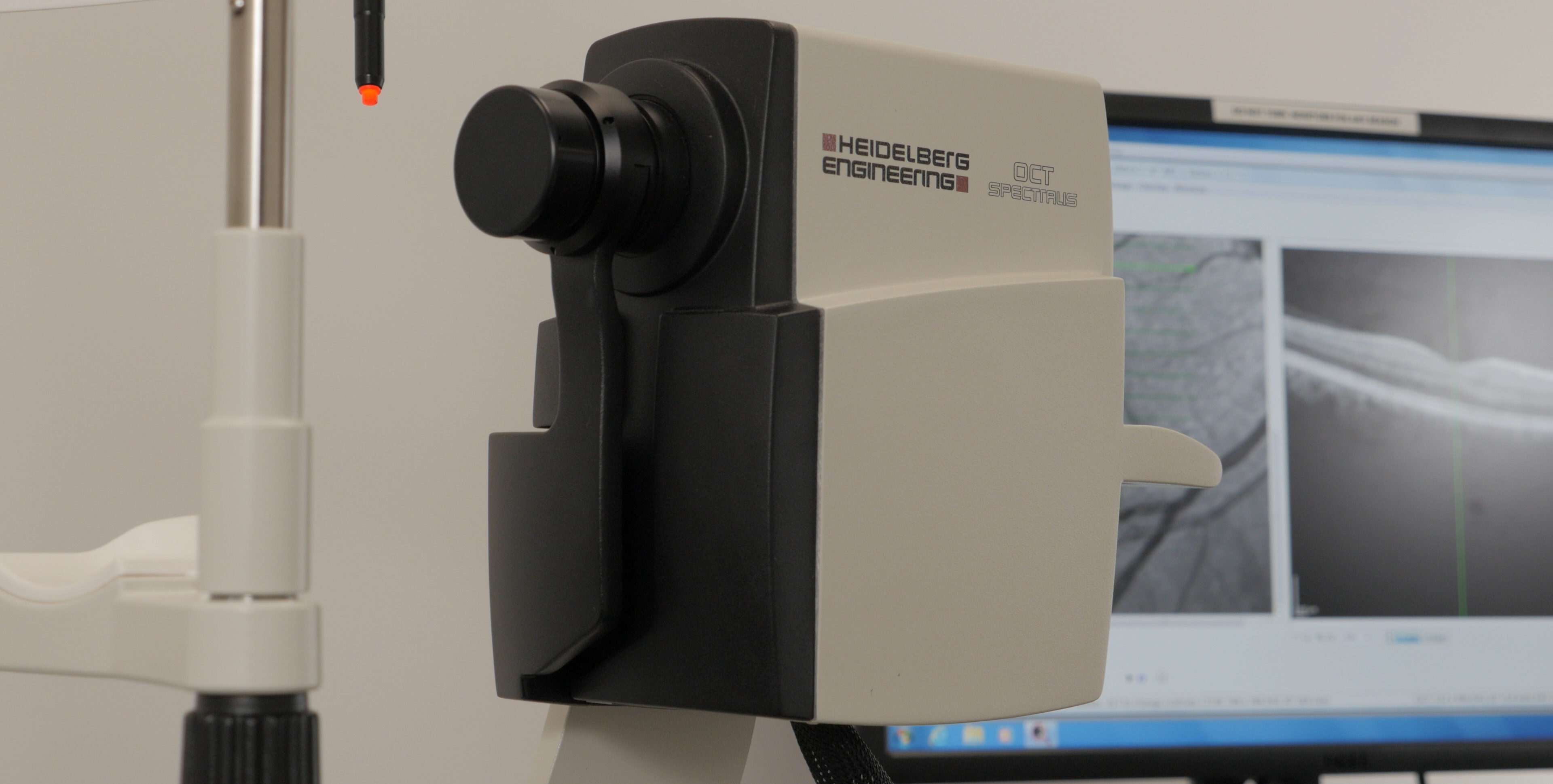
Optical Coherence Tomography (OCT) uses a non-invasive safe beam of light with special processing to look at the structures within the retina in very high detail. The anatomic features visible with OCT are much more detailed than that seen by normal examination. Seeing the retina from this cross sectional viewpoint provides a powerful way to detect, diagnose, and treat retinal disease.
Pathological changes associated with age-related macular degeneration, diabetic macular edema, central serous chorioretinopathy, and retinal edema associated with retinal vein occlusions can be detected and monitored with this form of imaging. OCT imaging is crucial in diagnosing and following glaucoma with scans designed to calculate changes to the optic nerve and surrounding nerve fiber layer. OCT is also imperative in measuring deeper layers, such as the choroid, which can be affected in many ocular and systemic diseases.
The latest glaucoma diagnostics are available using the SPECTRALIS® Glaucoma Module which compares patients’ eyes to a reference database of normal eyes, noting even very small deviations. The precision of the SPECTRALIS AutoRescan function allows confident identification and monitoring of structural changes from visit to visit.


Tagged Technology
